Fibroids
Uterine fibroids are noncancerous growths of the uterus that often appear during childbearing years. They are also called leiomyomas or myomas. Uterine fibroids aren't associated with an increased risk of uterine cancer and seldom develop into cancer.
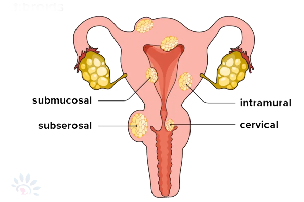
Fibroids range in size from small to big. one can have a single fibroid or multiple fibroids. In extreme cases, multiple fibroids can expand the uterus so much that it reaches the rib cage.

Causes:
Genetic changes:
Many fibroids contain changes in genes that differ from those in normal uterine muscle cells.
Hormones:
Estrogen and progesterone, two hormones that stimulate the development of the uterine lining during each menstrual cycle in preparation for pregnancy, appear to promote the growth of fibroids.
Fibroids contain more estrogen and progesterone receptors than normal uterine muscle cells do. Fibroids tend to shrink after menopause due to a decrease in hormone production.
Other growth factors:
Substances that help the body maintain tissues, such as insulin-like growth factors, may affect fibroid growth.
Extracellular matrix (ECM):
It is the material that makes cells stick together, like mortar between bricks. ECM is increased in fibroids and makes them fibrous. ECM also stores growth factors and causes biological changes in the cells themselves.
SYMPTOMS
Many women don't have any symptoms of uterine fibroids.
But some women may have symptoms of uterine fibroids like:
- Heavy menstrual bleeding
- Menstrual periods lasting more than a week
- Pelvic pressure or pain
- Frequent urination
- Difficulty emptying the bladder
- Constipation
- Backache or leg pains
Pregnancy and Fibroids
Fibroids usually don't interfere with getting pregnant. However, it's possible that fibroids — especially submucosal fibroids — could cause infertility or pregnancy loss.
Fibroids may also raise the risk of certain pregnancy complications, such as placental abruption, fetal growth restriction, and preterm delivery.
Diagnosis of uterine leiomyomas
It is generally made by comprehensive physical examination and clinical history. On physical exam, the most common finding is an enlarged uterus that is often irregular in shape. Confirmation of the clinical diagnosis is most easily done with ultrasonography.
Treatment
Surgery has traditionally been the gold standard for the treatment of uterine leiomyomas and has typically consisted of either hysterectomy or myomectomy. In recent years, a few clinical trials have evaluated the efficacy of orally administered medications for the management of leiomyoma-related symptoms.